The focus of our research is the development of extracorporeal blood purification techniques, in particular adsorption technologies in various applications and the supportive therapy of inflammation and sepsis.
In this context, we are working in the field of biocompatibility of biomaterials, on the influence of anticoagulation procedures on the blood compatibility of extracorporeal procedures, with the further development of blood compatible surfaces, furthermore with cell culture models to study the activation of the endothelium under septic conditions, as well as with the role and biological activity of extracellular vesicles.
Excellent infrastructure and a constantly evolving range of methods in the field of medical process engineering, cell culture and medical biochemistry as well as cooperation with clinical partners enable the transfer of research work from the basis into the application.

Infrastructure
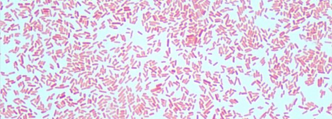
Our center has a laboratory area of approx. 500 m² with equipment in the fields of biochemistry, medical process engineering, microbiology and cell culture. The laboratories are equipped for work on biological safety levels 1 and 2. Furthermore, a Core Facility with advanced imaging equipment is available at Campus Krems.
- Autoanalyzer for clinical parameters (cobas/Roche C311)
- Chromatographic separation methods (Column Chromatography, HPLC)
- Flow Cytometer (Cytoflex LX) and Cell Sorter (FACSymphony S6)
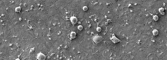
- Elektron microscope with EDX (Hitachi FlexSEM 1000) and Sputter-Coater
- Fluorescence microscopy (Olympus BX51)
- Gel electrophoresis and gel documentation system (BioRad ChemiDocTM XRS)
- Coagulation analyzer (Sysmex CA-560)
- Luminex Bead Array
- Measuring device for blood count (Sysmex XN 330)
- PCR (Roche Light Cycler 96) and MagNA Pure
- Cell culture models (ibidi µ-slide microfluidic system)
- Confocal Microscopy (Leica)
- Nanoparticle Tracking Analysis (ZetaView TWIN)
We have access to the following methods via our cooperation partners:
- Atomic Force Microscopy
- Cell Sorter
- Cryo-Electron microscopy (Tecnai F30 Polara transmission electron microscope)
- Imaging Flow Cytometry (ImageStreamx MkII cytometer, Amnis)
Methods
- Size exclusion chromatography
- Quantification of cytokines und mediators: ELISA, Luminex Bead Array
- Blood count, coagulation analysis, thrombin generation
- Characterization of adsorbents for extracorporeal blood purification:
- Adsorption capacity
- Adsorptions kinetics
- Pore structure
- Surface characterization
- Size exclusion
- Charakterisation of extracellular vesicles
- Nanoparticle Tracking Analysis
- Flow cytometry
- Imaging Flow Cytometry
- Blood material interactions
- Activation of the coagulation system
- Immunomodulation
- Charakterisation of membranes:
- Sieving coefficient
- Transmembrane pressure
- (Ultra)Filtrations rates
- Impact of the anticoagulation on blood compatibility
- Electrophoresis & Western Blotting
- Endotoxin quantification with the Limulus-Amebocyte-Lysate Test
- Characterization of polymers regarding blood cell activation
- Cell culture models for the characterization of the endothelial activation under septic conditions
Tags